Current Neuro-Ophthalmology Research Projects
The Photo-Blink Reflex
We are studying the dynamics of the photic blink reflex to objectively measure:
1. Afferent light sensitivity in the retina and trigeminal network
2. Efferent eyelid dysfunction in different disorders.
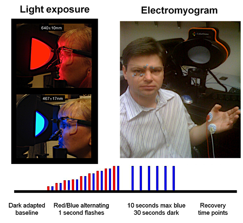
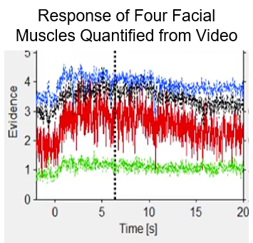
Quantitation of Facial Responses to Light
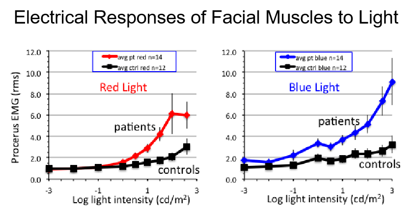
Facial Responses to Light Are Exaggerated in Light Sensitive Patients
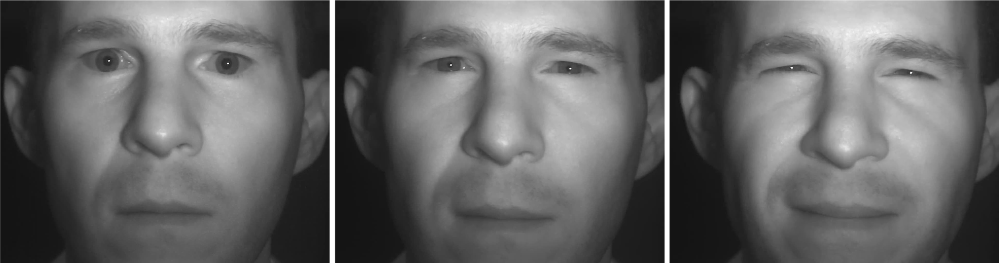
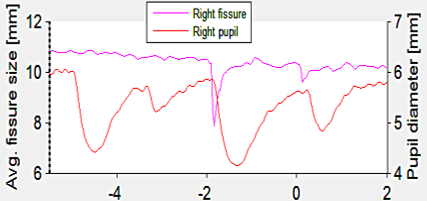
Real-Time 3D Facial Tracking
Automated pupil, eyelid and facial feature responses to light from an accessorized iPad.
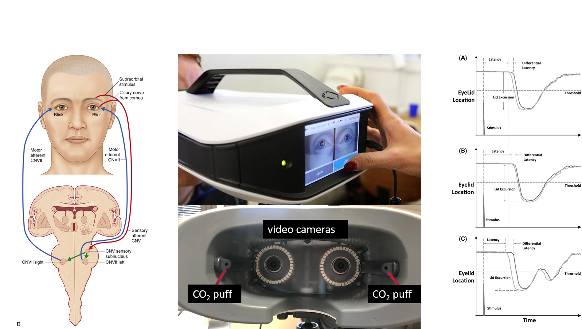
Corneal Blink Reflex – C02
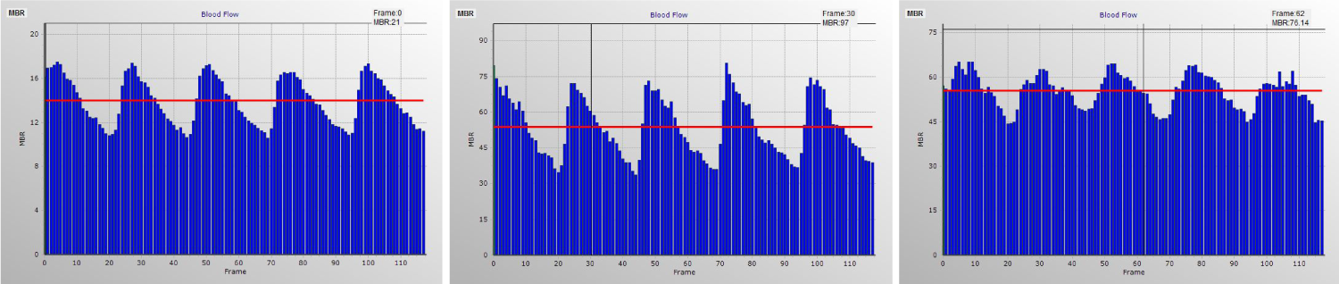
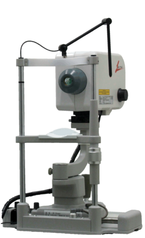
Laser Speckle Flowgraphy
Introduction to Laser Speckle Flowgraphy (LSFG)
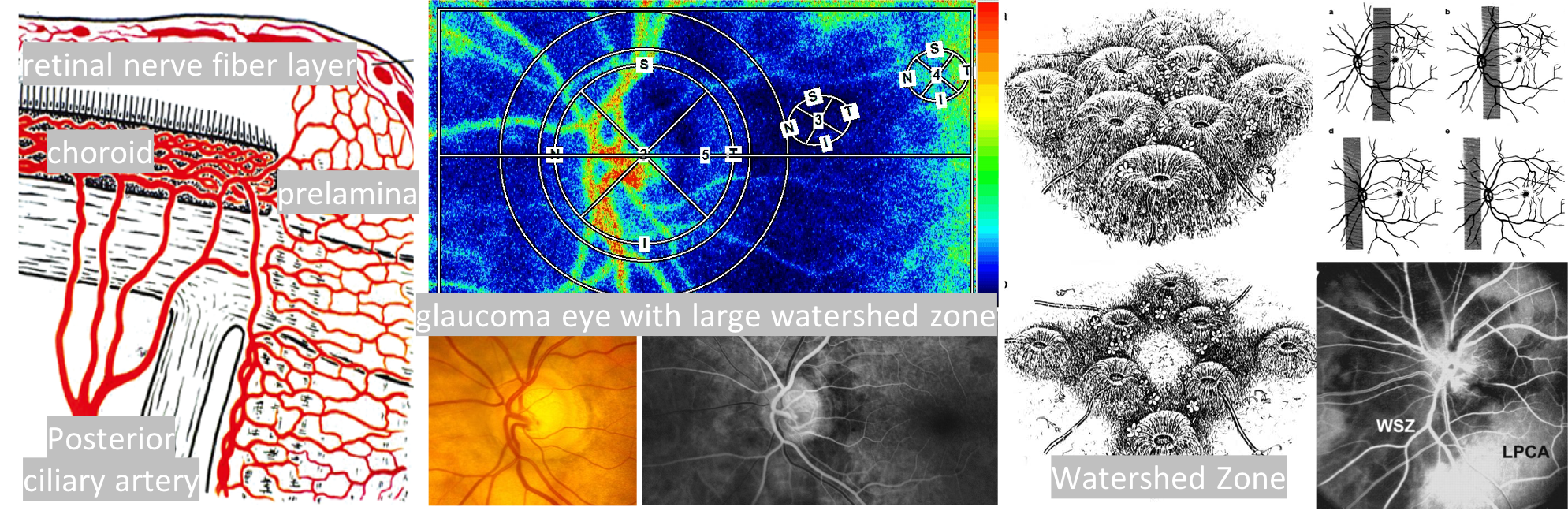
Blood Flow Quantified Simultaneously in the Retina, Optic Nerve and Choroid
This motion picture demonstrates the blood flow change in retinal vessels, tissue of optic nerve head and the choroid of human eye. You note the blood flow changes periodically being synchronized with the heart beats. From this time-varying map you may realize the LSFG’s potential to become a useful tool, not only for diagnoses of various eye diseases, but also for studying the functions of cardiovascular system.
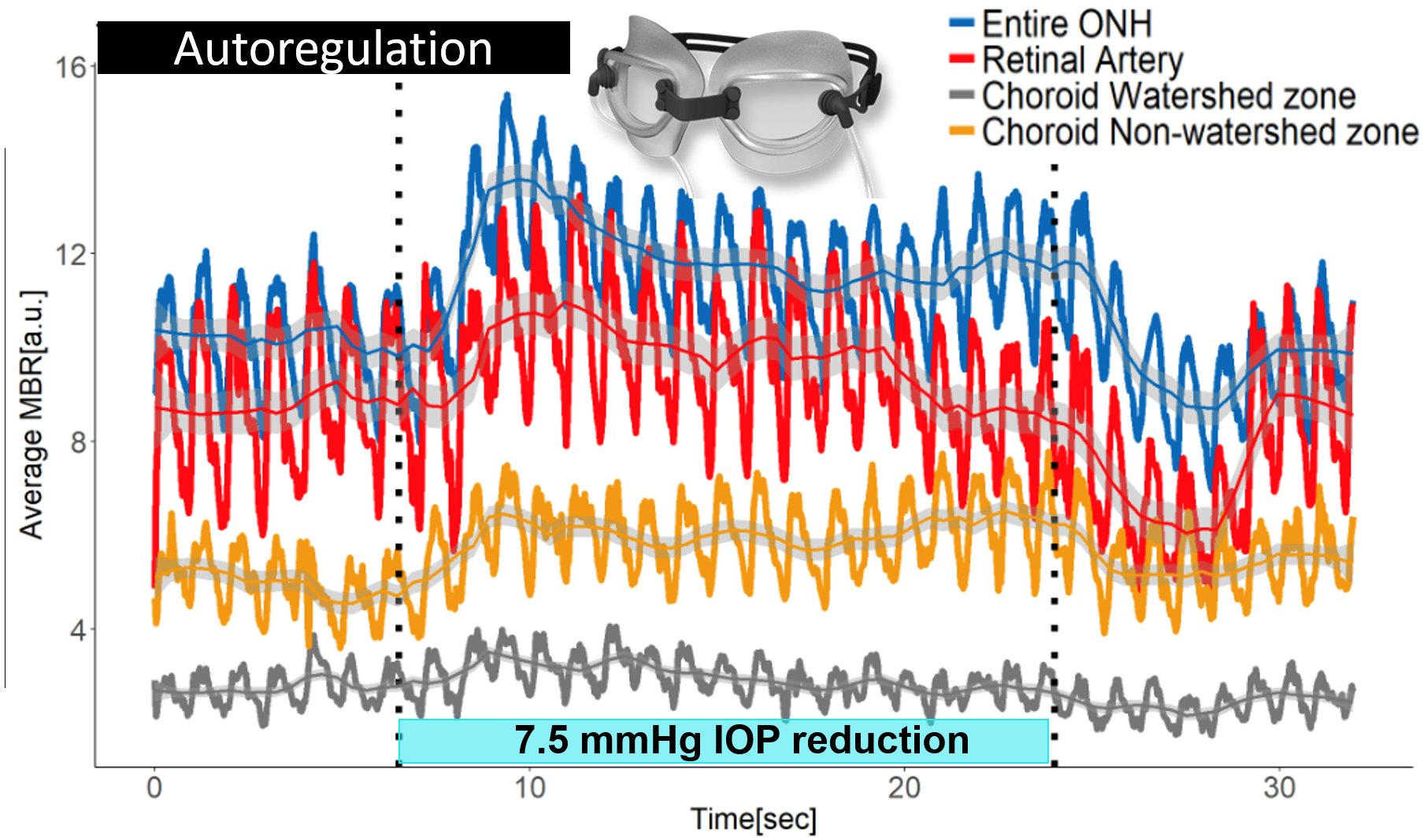
Use of Ocular Blood Flow to Test New Treatments
Laser speckle flowgraphy can be used to discover new treatments for improving blood flow to the eye and brain.
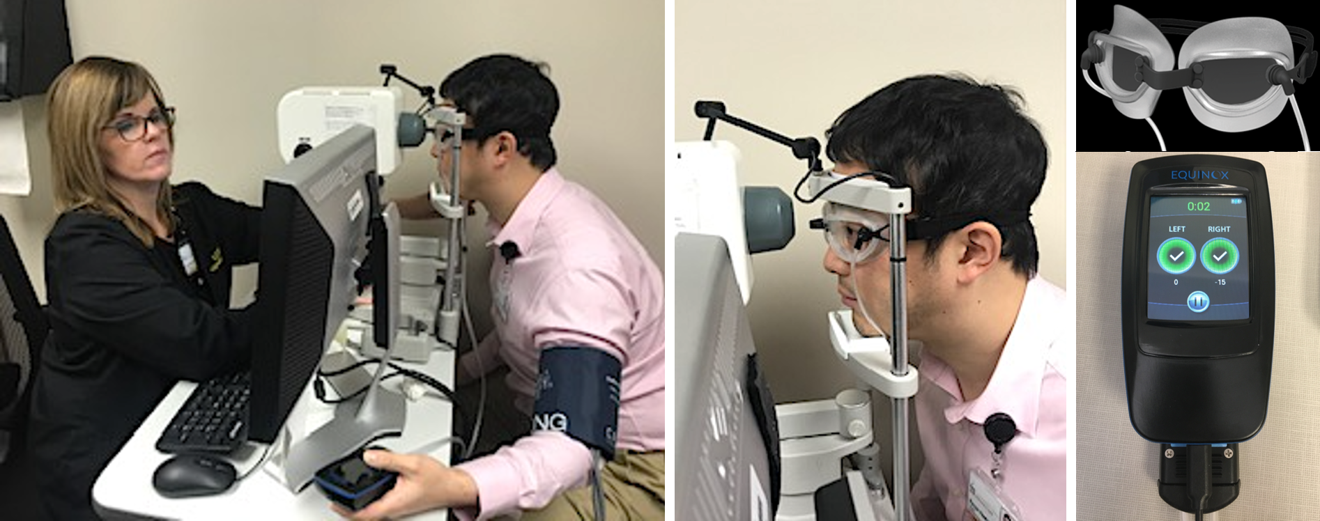
Measurement of ocular circulation with LSFG-NAVI while IOP acutely lowered using vacuum goggles
Light Flicker Evoked Increases in Ocular Blood Flow
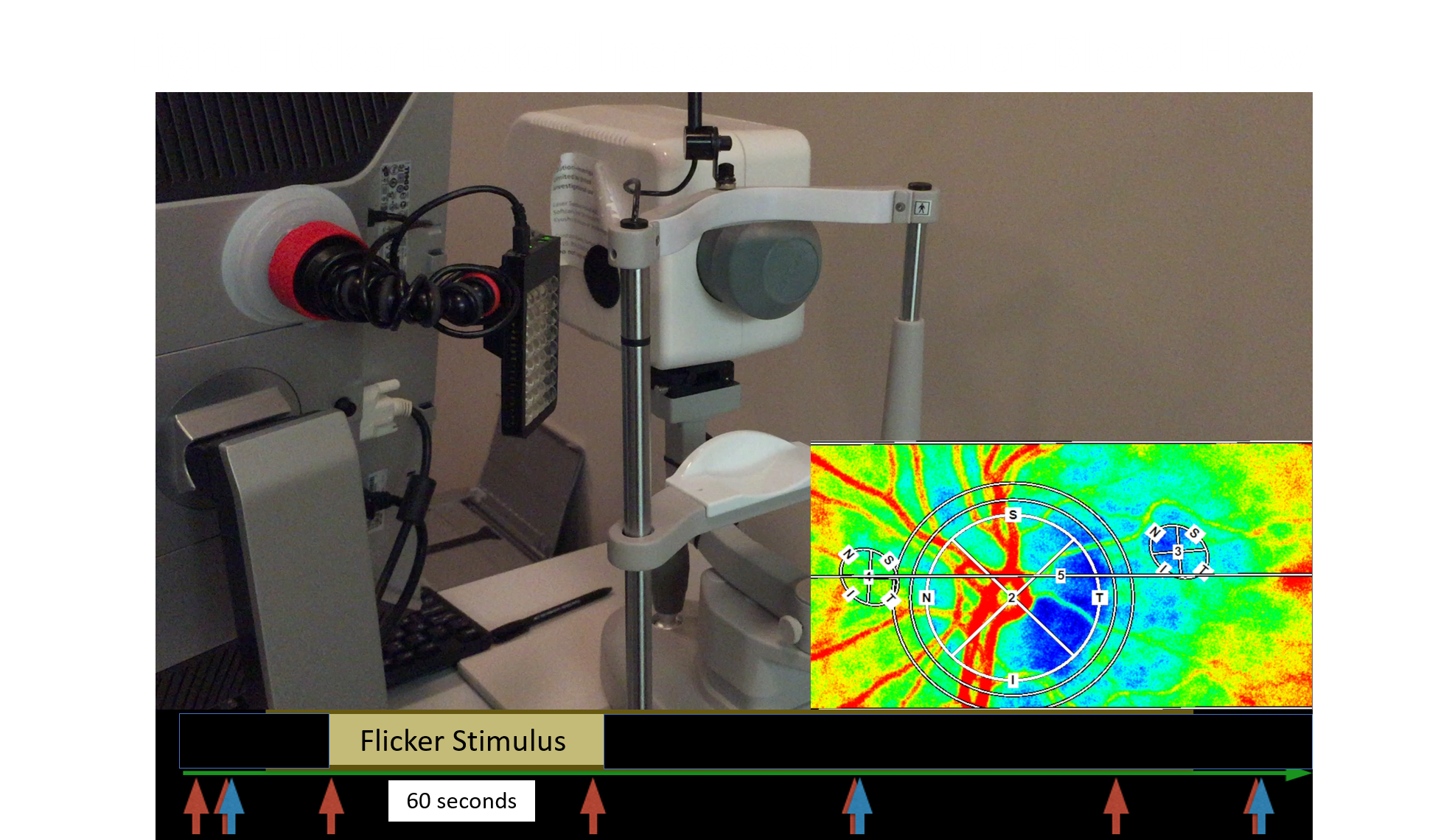
Blood Flow Changes in Response to Light Flicker
Optic Nerve Head Blood Flow Increases in Response to 10 Hz Light Flicker
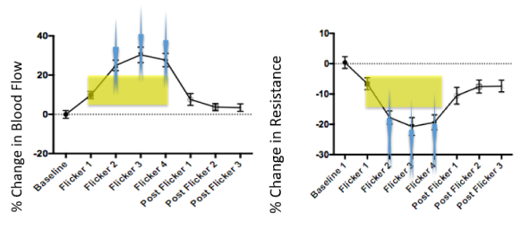
Perfusion Pressure/ Flow = Resistance
Perfusion Pressure = MAP-IOP
Ocular Perfusion Pressure =2/3 MAP-IOP
Laser Speckle Measurement in response to Light Flicker
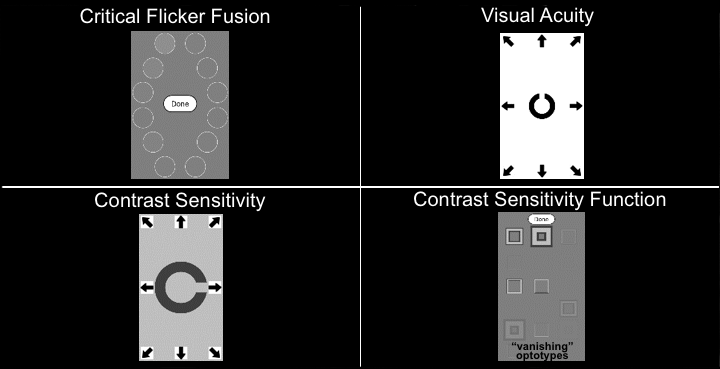
Smartphone Test of Visual Function